Respiratory System Homework Help
Respiration is one of the most important functions for the survival of human beings on the earth. The main function of respiratory system is to supply oxygen to our body thorough blood.
When we take in oxygen and release out carbon dioxide, several organ of our body comes into play. Starting from the nose, trachea, lungs and diaphragm, these entire organ serve as an important function for respiration.
Nose: nose is an important organ for respiration because the maximum air we breathe comes from nose only. Hence, it is the first organ that serves in different ways to give a better output. The hairs in the nose trap any contaminants in the air thus acting as filter and purifying the air. Nose also acts as the temperature calming device so that the air that is taken in, is made as per the suited temperature for the body i.e. if the air is too cold then nostril will make it warmer and if is too hot, nostril will make it cooler as per the body temperature. Above all, nostrils secrete mucous, that lubricates the nose keeping it moist. Therefore, the air passing from the nose will be filtered and purified before reaching the lungs.
Trachea: it is another important organ of respiratory system. It is a tube of 11cm long that extends from larynx to the fifth vertebrae in the chest cavity. The inner membrane of trachea is covered with tiny hairs that catch small hairs and removes it. It is surrounded with c shaped ring that protects it from any damage.
Histology of trachea: The tracheal walls from the deep to the superficial consist of: 1. Mucosa, 2. Sub mucosa, 3. Hyaline cartilage, 4. Adventitia.
1. Mucosa: It consists of Pseudo stratified columnar epithelium which is ciliated and below this is an underlying layer of lamina propria that contains elastic and reticular fiber.
2. Sub mucosa: it consists of sera mucous glands and their duct.
3. Hyaline cartilage: about 16-20 resembling the letter C is arranged one after the other. The open part of the C- shaped cartilage ring faces posterior towards the esophagus and contains the fibro muscular membrane.
Lungs: it is enclosed and protected by a double layered membrane called the pleural membrane. The superficial layer is called parietal pleura that line the wall of thoracic cavity and the deep player is called visceral pleura that covers the lungs. Between the visceral and parietal pleura is the small space called pleural cavity.

Respiratory System Assignment Help
Histology of lung:
1. The alveoli in the lungs are the functional unit of the lungs.
2. The wall of the alveoli consists of 2 types of alveolar epithelial cells, type 1 and type 2.
a. Type 1: they are most numerous and represent about 90% of the total alveolar surface. They are simple squamous epithelial cell, which are thin, which facilitates exchange of gases through the alveolar capillary membrane.
Type 2: alveolar cell also called septal cells. They are fewer in number and found between type-1 alveolar cells. The type- 1 are the main sites of gas exchange whereas the type 2 are rounded and cuboidal epithelial with free surface containing micro villi that secrete alveolar fluid. This fluid contains surfactant (prevent collapse of lung).
Surfactant is a complex mixture of phospholipids and lipoproteins. It lowers the surface tension of alveolar fluid, which reduces the alveoli to collapse and thus maintain the patency or their functionality. The alveolar wall also contains the alveolar macrophages which phagocyte fine dust particle.
3. The exchange of CO2 and O2 between the alveoli and the blood takes place by diffusion through the respiratory membrane or alveolar capillary membrane.
The respiratory membrane consists of 4 layers: extending from the alveolar air space to the blood plasma.
a. Alveolar Wall- which consist of type 1 and 2 and of macrophages.
b. Below the alveolar wall an epithelial basement membrane.
c. Capillary basement membrane and
d. Capillary endothelium.
Pulmonary ventilation:
The process of gas exchange in the body called respiration has 3 basic steps:
1. The respiratory minute volume or breathing: the amount of air inspired or expired per minute that is equal to the tidal volume multiplied by the respiratory rate (in a minute).
2. Pulmonary respiration: it is the exchange of gases between the alveoli of the lungs and the blood in pulmonary capillaries across the respiratory membrane.
3. Internal/ tissue respiration: it is the exchange of gases between bloods in systemic capillaries. In this process oxygen enters from the capillaries into the tissue and co2 enters from the tissues into the capillaries.
Within cells the metabolic reactions that consume oxygen and give out co2 during the production of ATP is termed as cellular respiration.
Therefore, Respiration= Inspiration + expiration
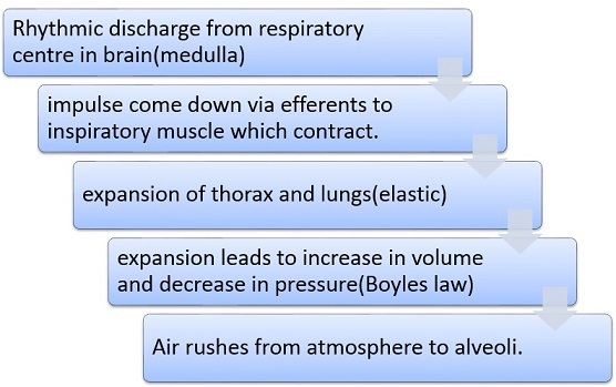
Inspiration:

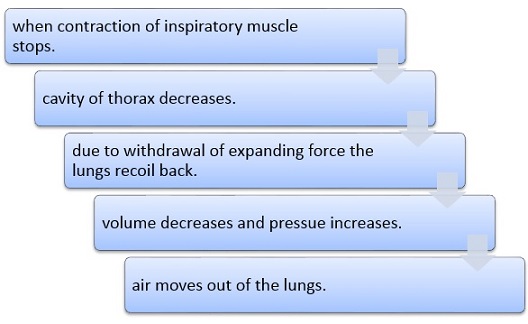
Expiration:

Inspiratory muscle:
- Diaphragm: it is supplied by phrenic nerve. It is the main muscle responsible for 70% of inspiration. It is dome shaped muscle which flattens on contraction from the floor of thoracic cavity.
- External intercostals: when these muscle contract, they move upward and outward, and is responsible for 25-30% inspiration.
- Accessory Inspiratory muscle: during forced inspiration.
- Sternocleidomastois: This elevates sternum muscle.
- Scalen: raises the first two ribs.
- Pectoralis minor: elevates the third through fifth ribs.
No muscles are required for normal expiration which is entirely due to recoil of lungs.
Muscles due to forced expiration:
1. Internal intercostals: the contraction of this depresses the ribs.
2. Abdominal muscles: it causes upward moment of viscera and pushes the diaphragm upward.
Factors affecting the pulmonary ventilation:
1. Compliance of the lung: it is defined as the change in lungs volume per unit change in alveolar pressure and is expressed as liter/cm of H2O.
2. Surface tension of alveolar fluid: surfactants reduce the surface tension and prevent the collapse of lung. Deficiency of surfactant in premature infants causes respiratory distress syndrome.
Some of the important terms:
1. Intrapleural pressure: before start of inspiration, the pressure in the intrapleural space is about 756 mm Hg. As the diaphragm and the external intercostals muscle contracts the volume of intrapleural cavity also increases which causes further fall of intrapleural pressure to about 754 mmHg.
2. Intrapulmonic pressure: with expansion of the lungs, the alveolar pressure falls below the atmospheric pressure to 758 mmHg and air rushes in from atmosphere to lungs.
During expiration the lungs recoils back as the pressure increases to 762 mmHg and air rushes from lungs to the atmosphere.
Respiratory volume and capacities: it is measured with the help of Spirometer and the record is called spirogram. The inhalation is recorded as an upward deflection and exhalation is recorded as a downward deflection.
- Tidal volume (TV): is the volume of air inspired or expired during each normal respiration
- Inspiration reserve volume (IRV): is the maxi amount of air that can be inspired just after normal inspiration.
- Inspiratory capacity (IC): is the maximum amount of air that can be inspired just after normal expiration.
- Vital capacity (VC): is the maximum amount if air that can expire by force full effort after maximum inspiration.
- Residual volume (RV): is the amount of air that can be expired by force full effort after maximum expiration.
- Functional residual capacity (FRC): is the amount of air that in the lungs after normal expiration.
- Total lung capacity (TLC): is the total amount of air that in the lung after maximum inspiration.
- Timed vital capacity (TVC): measured in forced expiratory volume. It is measured on a fast moving Spirometer is recorded in 1 second, 2 second, 3 second (FEV1, FEV2, FEV3) normally all the volume of air can be expired in less than 4 second. In some disease like COPD< chronic obstructive pulmonary disease> which increases the air ways resistance, reduce FEV1.
- Pulmonary ventilation (PV): is the amount of air inspired or expired per minute. = (T.V*RR=500*12-15=6-7.5 L/MINUTE).
- Dead space: anatomical (respiratory dead space) it is volume of conducting air ways from nose to mouth up to terminal bronchioles where exchange of gases does not takes place.
Alveolar ventilation: is the amount of air ventilating the alveoli per minute which is equal to:
= (tidal volume-dead space) XRR
= (500-150) *12-15
=4-5 l/minute.
Therefore, overall respiratory system serves an important function in the body keeping us alive, and helping us live a healthy life. Though the system seems simple and easy but the procedure is far more complex than anyone can even expect. Therefore to keep our respiratory system proper functioning and free of any diseases, we must be careful and remain away from polluted places and ignore any bad habits, thus live in healthy atmosphere.
Topics In Biology
- Anatomy
- Biochemistry
- Bioinformatics
- Cell Biology
- Digestive system
- Ecology
- Entomology
- Epidemiology
- Immunology
- Microbiology
- Molecular Biology
- Paleontology
- Respiratory System Homework Help
- Recombinant DNA Technology
- Zoology
- Biophysics
- Genetics
- Physiology
- Nervous system

